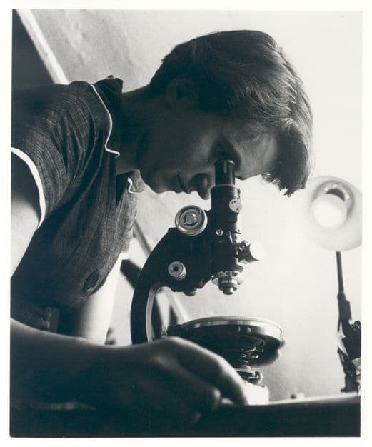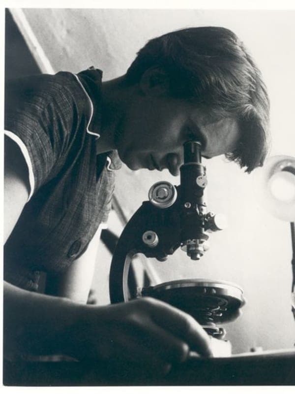
On 6 May 1952, in a basement laboratory beneath the chemistry building at King's College London, Rosalind Franklin and her graduate student Raymond Gosling exposed a fiber of DNA to X-rays1 for roughly one hundred hours. The image that came back — the fifty-first diffraction pattern Franklin had shot of DNA — showed a crisp, unmistakable X at its center.2 That cross shape encoded the geometry of the double helix: its dimensions, pitch, and the offset between its two strands.2 Franklin recognized it immediately, noting in her records that the B form of DNA had a helical structure.3 The photograph would become the most consequential X-ray image in the history of biology, and Franklin had built the instrument to capture it herself.4
Early Life
Franklin spent the four years between 1947 and 1951 in Paris, working under Jacques Mering at the Laboratoire Central des Services Chimiques de l'État5 — a government chemistry institute where, as one colleague noted, women were engaged as equals5. She had arrived with a Cambridge PhD in physical chemistry and a thesis on the porosity of coal; she left as an accomplished X-ray crystallographer. At the Laboratoire Central she carried out powder diffraction on amorphous carbons, distinguishing the ones that graphitize when heated to extreme temperatures from those that form a rigid, finely porous mass — a classification that proved foundational for the coking industry and the development of heat-resistant materials.6 By the time she returned to England in 1951, she had published five papers on carbon structure5, held an international reputation among coal chemists, and mastered a technique she would spend the rest of her life applying to progressively harder problems.
Career
When Franklin joined King's College's MRC (Medical Research Council) Biophysics Unit in January 1951, almost nothing precise was known about the physical structure of DNA. She attacked the problem the way she had attacked coal: by controlling experimental conditions until the data became unambiguous. By carefully regulating the water content of DNA fibers, she established that the molecule existed in two distinct forms, A and B, each with a different X-ray signature.7 She designed and modified a micro-camera to capture sharper diffraction patterns than any produced before.4 Photo 51, taken in May 1952, encoded the key geometric facts of the B form.
Franklin published her findings in the April 1953 issue of Nature, in the same issue in which Watson and Crick announced their double-helix model — a model her data corroborated. What she almost certainly did not know at the time was that her unpublished results, including the measurements in her MRC progress report, had been seen by Watson months earlier without her knowledge. In their paper, Watson and Crick did not credit Franklin for supplying that evidence or for Photo 51; they noted only that they had been stimulated by knowledge of the "general nature" of the unpublished results of Wilkins, Franklin, and their co-workers. When Watson, Crick, and Wilkins received the Nobel Prize in Physiology or Medicine in 1962, neither Watson nor Crick mentioned Franklin in their Nobel lectures; Wilkins referred only to her having made "valuable contributions to the X-ray analysis." It was not until decades after her death that Watson and Crick, in separate retrospective accounts, acknowledged the indispensability of her data — Crick allowing that all the really relevant experimental work on the diffraction patterns had come from her lab, Watson conceding that the discovery would not have been possible without it.
Franklin left King's in March 1953 and moved to J. D. Bernal's crystallography laboratory at Birkbeck College.9 She spent the next five years building the field of structural virology. Beginning with tobacco mosaic virus — the first organism ever identified as a viral pathogen — she obtained X-ray diffraction patterns of unprecedented clarity and used them to show that TMV's RNA was wound in a helical groove along the inner wall of the virus's protein shell.10 From there she expanded to turnip yellow mosaic, tomato bushy stunt, and other plant viruses, preparing more than a dozen papers for publication in 1956 and 1957 alone.10 After a 1956 visit to the University of California, Berkeley, she turned her attention to poliovirus11: still a major killer, only recently targeted by Salk's vaccine. She applied for and received a three-year grant from the U.S. National Institutes of Health — the largest external research fund Birkbeck had ever received — to decipher the crystal structure of the virus.12 She was by then being invited to speak at conferences across Europe and the United States, frequently as the only woman on the program.7
Legacy
Franklin was diagnosed with ovarian cancer in the autumn of 1956.13 She underwent multiple surgeries and several periods of remission, and she continued working throughout, pressing forward on the poliovirus crystal structure until her health made it impossible. She died in London on 16 April 1958, thirty-seven years old.13 In the obituary he wrote for Nature, J. D. Bernal described her photographs as 'among the most beautiful x-ray photographs of any substance ever taken.'14 Her colleague Aaron Klug, who took over her research group after her death, published the poliovirus structure the following year with John Finch, dedicating the paper to her memory.6 Klug was awarded the 1982 Nobel Prize in Chemistry for his work on the structure of nucleic acid-protein complexes — work he began with Franklin at Birkbeck.12 Throughout her sixteen-year career Franklin had published 19 papers on coals and carbons, 5 on DNA, and 21 on viruses.13 At the 1958 Brussels World's Fair, a five-foot model of the tobacco mosaic virus that Franklin had built — using table tennis balls and plastic bicycle handlebar grips — went on display at the International Science Pavilion on 17 April 1958, the day after she died.11